Introduction
At the cutting edge of ophthalmic innovation, Nishka Research presents a pioneering approach to corneal lenticule analysis by synergizing Scanning Electron Microscopy (SEM) and Optical Microscopy (OM) techniques. The extensive analysis is pivotal for understanding the refractive surgery outcomes, particularly in procedures like ReLEx SMILE.
The Challenge
The accuracy in corneal lenticule extraction and its transplantation is pivotal for the success of refractive surgeries. Conventional analytical methods will have limitations in providing the microscopic characterization required for optimizing surgical precision. The challenge lies in achieving a holistic approach of the lenticule's morphology and structural integrity.
Nishka Research's Integrated Approach
Combining SEM and OM Techniques: By integrating SEM's high magnification and high resolution images with the broader contextual insights of optical microscopy images, the data obtained will be more trustworthy and reliable. Nishka Research has developed a comprehensive methodology for Precision and Integrity evaluation of corneal lenticule. This dual approach enables unparalleled detail and accuracy in our analyses.
Methodologies adopted:
Results obtained
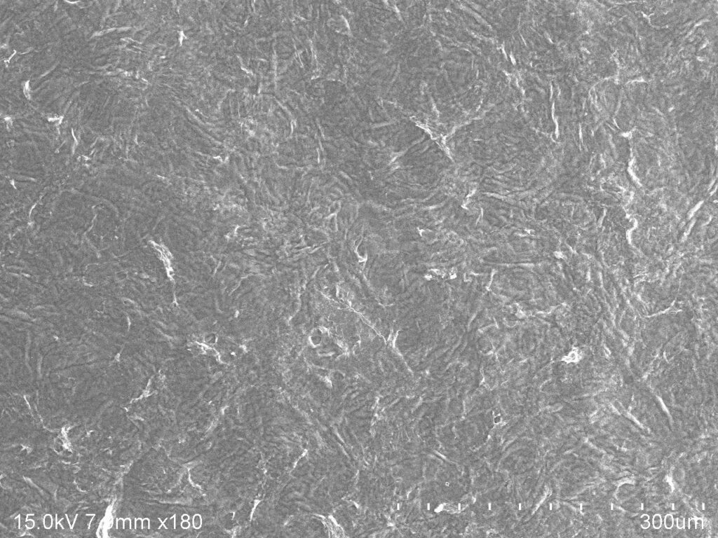
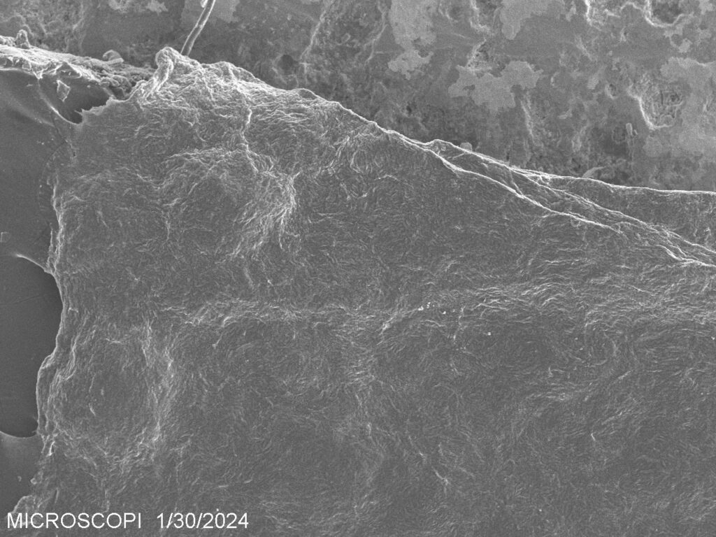
Integrated Insights: The combined application of SEM and OM provided a complete view of the corneal lenticule's quality. SEM offered detailed images of surface smoothness and edge sharpness, while OM contributed to understanding the lenticule's uniformity and macroscopic integrity.
Enhanced Surgical Precision: This integrative method of analysis has noteworthy implications for improving the precision of lenticule creation and transplantation, potentially reducing post-surgery complications and enhancing patient compliance.
Conclusion
The innovative approach of integrating Scanning Electron microscopy and Optical Microscopy at Nishka Research represents a leap forward in corneal lenticule characterization. The present case study not only showcases our capability to provide detailed, accurate assessments but also emphasizes our commitment to supporting the next generation of ophthalmic surgery techniques.
Learn more about the impact of Nishka Research's analytical services on ophthalmic surgery and beyond. Explore our services and further case studies to see how we're driving forward scientific discovery and healthcare solutions.
Need help or Have a question?
Nishka Research Pvt. Ltd.
Regus Business Center,
4th floor, Gumidelli Commercial Complex,
1-10-39 to 44, Old Airport Road, Begumpet,
Hyderabad-500016
India
Phone : +91 40 29303155
Mobile : +91 7842798518

Left us a star review


Great work done by nishka research. Thank you so much for your kind Co operation


A very good lab delivering acurate results. I must also mention about the very cordial staff that they have.

I am a PhD scholar from Kaunas University at Lithuania, Europe. I have given a sample to perform SEM analysis of the organic tissue. The results are highly satisfactory and impressive. The service was really excellent, on time( even before the time schedule), consumer friendly. cost to the academic samples are very less comparing with others. Once again thanks to Nishka for providing me the service ontime as I am really in urgency...


Submitted the samples for my phd work . Great work and reports submited with 2 days of time . Also the image clarity and all data are clear .


Thank you for your services.. Looking forward for more collaboration with your labs for my future projects..